Home
/ Microcalcifications Thyroid Nodule : Us Features Of Thyroid Malignancy Pearls And Pitfalls Radiographics, Benign nodules can be hypoechoic;
Microcalcifications Thyroid Nodule : Us Features Of Thyroid Malignancy Pearls And Pitfalls Radiographics, Benign nodules can be hypoechoic;
Microcalcifications Thyroid Nodule : Us Features Of Thyroid Malignancy Pearls And Pitfalls Radiographics, Benign nodules can be hypoechoic;. One of the most important ultrasound features of cancer is the presence of calcifications, especially microcalcifications, in a thyroid nodule. If the thyroid demonstrates diffuse uptake compatible with chronic lymphocytic thyroiditis, further imaging or fna is not. 20,21 however, this finding has a low sensitivity (29% to 59%), since microcalcification is often not present in malignant nodules. If no other malignant features (e.g. Tsh measurement should be part of the initial workup in every patient with a thyroid nodule and be used as a guide for further management (fig.
Such nodules are a common occurrence in the general population and a frequent incidental finding on computed tomography (ct) and magnetic resonance imaging (mri). Published guidelines recommend endocrinology consultation and biopsy. If no other malignant features (e.g. The presence of microcalcifications on an ultrasound is felt to be highly suggestive of thyroid cancer. In contrast, other studies have shown that ultrasound features such as coarse calcifications, more tall than wide, irregular borders and increased blood flow within the.
Https Encrypted Tbn0 Gstatic Com Images Q Tbn And9gcrnhpgd3eqf8ozznfklfwyijbnsfafbzasg2s6 Fx1uuftbhcau Usqp Cau from An incidental finding of focal fdg uptake in a >1 cm thyroid nodule is concerning and fna is warranted. This study suggests that ultrasound features of microcalcifications, solid nodule and size larger than 2 cm can be used to identify patients at high risk for thyroid cancer. 20,21 however, this finding has a low sensitivity (29% to 59%), since microcalcification is often not present in malignant nodules. One of the most important ultrasound features of cancer is the presence of calcifications, especially microcalcifications, in a thyroid nodule. 25% (follicular and medullary) hyperechoic solid nodule: If the thyroid demonstrates diffuse uptake compatible with chronic lymphocytic thyroiditis, further imaging or fna is not. Tsh measurement should be part of the initial workup in every patient with a thyroid nodule and be used as a guide for further management (fig. Published guidelines recommend endocrinology consultation and biopsy.
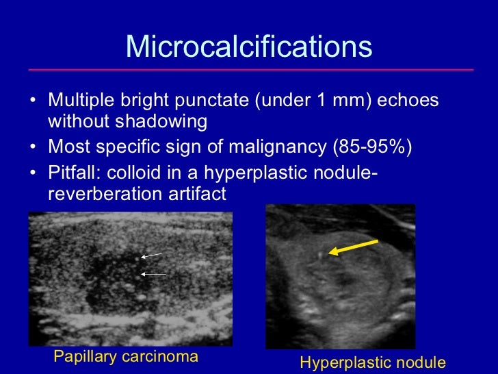
When present, fine, nonshadowing echogenic foci representing microcalcifications are highly indicative of papillary thyroid cancer, with a specificity of 95% (figures 4 and 5).
Such nodules are a common occurrence in the general population and a frequent incidental finding on computed tomography (ct) and magnetic resonance imaging (mri). The risk of malignancy in thyroid nodules increases as the serum tsh increases. 25% (follicular and medullary) hyperechoic solid nodule: In contrast, other studies have shown that ultrasound features such as coarse calcifications, more tall than wide, irregular borders and increased blood flow within the. The presence of microcalcifications on an ultrasound is felt to be highly suggestive of thyroid cancer. Nov 16, 2013 · background heterogeneous echogenicity of the thyroid gland has been associated with diffuse thyroid disease and benign and malignant nodules can coexist with diffuse thyroid disease. A thyroid nodule is a discrete lesion within the normal thyroid. Calcifications) then hypoechoic nodules are typically biopsied after reaching size criteria; When present, fine, nonshadowing echogenic foci representing microcalcifications are highly indicative of papillary thyroid cancer, with a specificity of 95% (figures 4 and 5). If <1 cm the nodule may be monitored similarly to a subcentimeter thyroid nodule with a high risk sonographic pattern; Underlying heterogeneous echogenicity might make it difficult to differentiate between benign and malignant nodules on us. If no other malignant features (e.g. 20,21 however, this finding has a low sensitivity (29% to 59%), since microcalcification is often not present in malignant nodules.
An incidental finding of focal fdg uptake in a >1 cm thyroid nodule is concerning and fna is warranted. The risk of malignancy in thyroid nodules increases as the serum tsh increases. One of the most important ultrasound features of cancer is the presence of calcifications, especially microcalcifications, in a thyroid nodule. If the thyroid demonstrates diffuse uptake compatible with chronic lymphocytic thyroiditis, further imaging or fna is not. Published guidelines recommend endocrinology consultation and biopsy.
The Epidemic Of Thyroid Nodules Which Should Undergo Fine Needle Asp from image.slidesharecdn.com An incidental finding of focal fdg uptake in a >1 cm thyroid nodule is concerning and fna is warranted. 1,24,25 a normal or high tsh level should raise concerns for possible malignant potential of a nodule, whereas a low tsh is an indicator of benignity in. Thus, the aim of this study was to evaluate the influence of underlying thyroid. 25% (follicular and medullary) hyperechoic solid nodule: Nov 16, 2013 · background heterogeneous echogenicity of the thyroid gland has been associated with diffuse thyroid disease and benign and malignant nodules can coexist with diffuse thyroid disease. If the thyroid demonstrates diffuse uptake compatible with chronic lymphocytic thyroiditis, further imaging or fna is not. If no other malignant features (e.g. 5% chance of being malignant
In contrast, other studies have shown that ultrasound features such as coarse calcifications, more tall than wide, irregular borders and increased blood flow within the.
If <1 cm the nodule may be monitored similarly to a subcentimeter thyroid nodule with a high risk sonographic pattern; When present, fine, nonshadowing echogenic foci representing microcalcifications are highly indicative of papillary thyroid cancer, with a specificity of 95% (figures 4 and 5). 25% (follicular and medullary) hyperechoic solid nodule: 5% chance of being malignant This study suggests that ultrasound features of microcalcifications, solid nodule and size larger than 2 cm can be used to identify patients at high risk for thyroid cancer. Calcifications) then hypoechoic nodules are typically biopsied after reaching size criteria; Benign nodules can be hypoechoic; In contrast, other studies have shown that ultrasound features such as coarse calcifications, more tall than wide, irregular borders and increased blood flow within the. A thyroid nodule is a discrete lesion within the normal thyroid. If no other malignant features (e.g. Published guidelines recommend endocrinology consultation and biopsy. 1,24,25 a normal or high tsh level should raise concerns for possible malignant potential of a nodule, whereas a low tsh is an indicator of benignity in. Thus, the aim of this study was to evaluate the influence of underlying thyroid.
A thyroid nodule is a discrete lesion within the normal thyroid. When present, fine, nonshadowing echogenic foci representing microcalcifications are highly indicative of papillary thyroid cancer, with a specificity of 95% (figures 4 and 5). Calcifications) then hypoechoic nodules are typically biopsied after reaching size criteria; If the thyroid demonstrates diffuse uptake compatible with chronic lymphocytic thyroiditis, further imaging or fna is not. In contrast, other studies have shown that ultrasound features such as coarse calcifications, more tall than wide, irregular borders and increased blood flow within the.
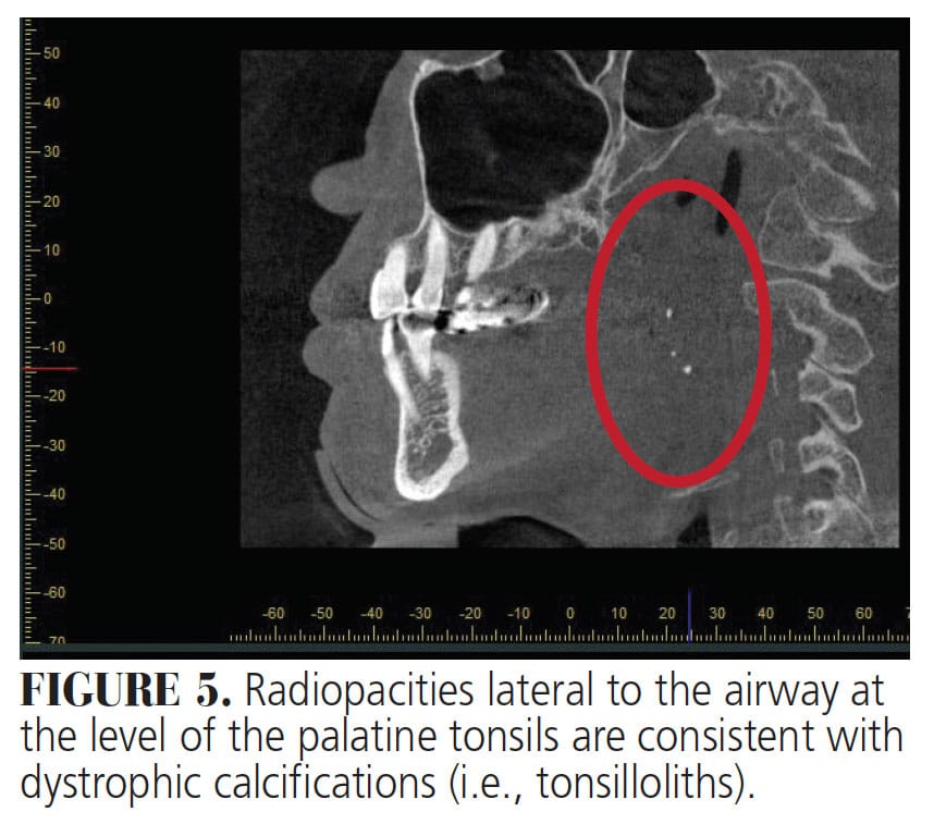
Diagnosing Incidental Thyroid Calcifications On Dental Images Decisions In Dentistry from decisionsindentistry.com Thus, the aim of this study was to evaluate the influence of underlying thyroid. In contrast, other studies have shown that ultrasound features such as coarse calcifications, more tall than wide, irregular borders and increased blood flow within the. If <1 cm the nodule may be monitored similarly to a subcentimeter thyroid nodule with a high risk sonographic pattern; This study suggests that ultrasound features of microcalcifications, solid nodule and size larger than 2 cm can be used to identify patients at high risk for thyroid cancer. The risk of malignancy in thyroid nodules increases as the serum tsh increases. If the thyroid demonstrates diffuse uptake compatible with chronic lymphocytic thyroiditis, further imaging or fna is not. 25% (follicular and medullary) hyperechoic solid nodule: Calcifications) then hypoechoic nodules are typically biopsied after reaching size criteria;
The presence of microcalcifications on an ultrasound is felt to be highly suggestive of thyroid cancer.
Such nodules are a common occurrence in the general population and a frequent incidental finding on computed tomography (ct) and magnetic resonance imaging (mri). If no other malignant features (e.g. The risk of malignancy in thyroid nodules increases as the serum tsh increases. Nearly all medullary thyroid carcinomas 3; 5% chance of being malignant An incidental finding of focal fdg uptake in a >1 cm thyroid nodule is concerning and fna is warranted. Nov 16, 2013 · background heterogeneous echogenicity of the thyroid gland has been associated with diffuse thyroid disease and benign and malignant nodules can coexist with diffuse thyroid disease. If <1 cm the nodule may be monitored similarly to a subcentimeter thyroid nodule with a high risk sonographic pattern; In contrast, other studies have shown that ultrasound features such as coarse calcifications, more tall than wide, irregular borders and increased blood flow within the. One of the most important ultrasound features of cancer is the presence of calcifications, especially microcalcifications, in a thyroid nodule. Nov 26, 2015 · introduction. Published guidelines recommend endocrinology consultation and biopsy. The presence of microcalcifications on an ultrasound is felt to be highly suggestive of thyroid cancer.